Who invented the microscope?
Short answer
The invention of the microscope is attributed to Dutch spectacle makers Hans and Zacharias Janssen around 1590. While they created the first compound microscope by placing two lenses in a tube, Antonie van Leeuwenhoek later revolutionized the field in the 1670s by developing high-powered single-lens microscopes capable of observing bacteria and protozoa.

On this page
Who actually invented the microscope?
The credit for the first compound microscope is widely given to Hans and Zacharias Janssen, a father-son team of spectacle makers in Middelburg, Netherlands. Around 1590, they discovered that placing two lenses inside a tube allowed them to magnify small objects significantly. Their device was relatively crude, offering only about 3x to 9x magnification, but it established the fundamental principle of compound optics that remains the basis for modern laboratory instruments.
However, the history of magnification is complex and involves several contributors. Galileo Galilei, the famous Italian astronomer, refined the design in 1609 to create an "occhiolino" or "little eye," which used a convex and a concave lens. While Galileo’s device was technically impressive, it was Antonie van Leeuwenhoek who truly unlocked the microscopic world. By the 1670s, he had mastered the art of grinding tiny, high-curvature glass lenses that could magnify objects up to 270 times, far surpassing the capabilities of early compound designs.
How did early microscopes work?
Early compound microscopes functioned by using two separate lens systems: an objective lens and an eyepiece. The objective lens gathered light from the specimen and created a magnified image inside the tube, which the eyepiece then magnified further for the observer's eye. This dual-stage magnification allowed for greater detail than a simple magnifying glass, though early glass quality often introduced significant distortions and chromatic aberrations.
Antonie van Leeuwenhoek’s approach was different because he utilized a single, powerful lens. His microscopes were small, handheld devices that required the user to place the specimen on a sharp needle point directly in front of the lens. This design minimized the light distortion found in compound microscopes of the era. To achieve such high magnification, he developed a unique technique for grinding and polishing glass beads, which allowed him to be the first human to document:
- Bacteria found in dental plaque.
- The movement of sperm cells.
- The structure of red blood cells.
- The complex anatomy of tiny insects.
What are the main types of microscopes?
Today, microscopes are categorized primarily by the source of illumination and the way they manipulate that light. Optical or light microscopes use visible light and glass lenses to magnify specimens, making them ideal for viewing living cells or thin tissue sections. Electron microscopes, by contrast, use beams of electrons to achieve much higher resolution, allowing scientists to see structures as small as individual atoms.
The most common types used in research and education include:
- Compound light microscopes: Standard for biology classrooms and clinical labs.
- Stereo microscopes: Used for dissecting or viewing larger, opaque objects in 3D.
- Scanning Electron Microscopes (SEM): Used to create detailed 3D surface images of specimens.
- Transmission Electron Microscopes (TEM): Used to view the internal molecular structure of samples.
Choosing the right type depends entirely on the desired level of magnification and whether the specimen needs to be alive or dead during observation.
How do you use a microscope properly?
Using a compound microscope requires a systematic approach to ensure the image is clear and the specimen is not damaged. Start by placing your slide on the stage and securing it with stage clips. Always begin with the lowest power objective lens to locate your specimen, as this provides a wider field of view and makes focusing much easier.
Follow these steps to ensure a successful viewing session:
- Place the slide on the stage and center the specimen over the light hole.
- Use the coarse adjustment knob to bring the specimen into rough focus while looking from the side to avoid hitting the slide.
- Look through the eyepiece and slowly turn the fine adjustment knob until the image is crisp.
- Once focused, rotate the nosepiece to a higher power objective if more detail is required.
- Adjust the diaphragm to control the amount of light passing through the slide for optimal contrast.
Common mistakes when using a microscope
One of the most frequent errors beginners make is using the coarse adjustment knob while on a high-power objective lens. This can cause the lens to strike the slide, potentially shattering the glass and damaging the expensive objective lens. Always use the coarse adjustment only on the lowest power setting, and rely exclusively on the fine adjustment knob for higher magnifications.
Another common issue is improper lighting, which often results in a washed-out or dark image. Users frequently forget to adjust the condenser or the iris diaphragm, which regulates the aperture of the light beam. If the image is too bright, the contrast will be lost; if it is too dark, the details will be obscured. Finally, failing to clean the lenses with specialized lens paper can leave fingerprints or dust, which will appear as dark, distracting spots in your field of view. Always handle lenses with care to maintain optical clarity for your observations.
Key facts
- Hans and Zacharias Janssen, a Dutch father-son team of spectacle makers, are widely credited with creating the first compound microscope around 1590.
- Antonie van Leeuwenhoek, a Dutch scientist, significantly improved microscope design in the 1670s by using high-quality single lenses to achieve magnifications of up to 270x.
- A compound microscope uses two or more lenses to magnify an image, while a simple microscope uses only one lens.
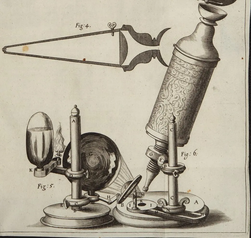
- Robert Hooke published "Micrographia" in 1665, introducing the term "cell" after observing thin slices of cork under a microscope.
- When using a microscope, always start with the lowest magnification objective lens to make finding your specimen easier.
- Avoid touching the glass surface of lenses with your fingers, as skin oils can permanently damage the delicate optical coatings.
The bottom line
The invention of the microscope was not the work of one individual, but rather an evolution of optical discoveries by Dutch spectacle makers and scientists. While the Janssens pioneered the compound design in the late 16th century, figures like Leeuwenhoek and Hooke transformed it into a vital scientific tool. Today, these instruments remain essential for understanding the microscopic structures that define our biological and physical world.
People also ask
What is the difference between a simple and a compound microscope?
A simple microscope uses a single convex lens, similar to a magnifying glass, to enlarge an object. A compound microscope uses a two-lens system—an objective lens and an eyepiece—to provide much higher magnification and resolution.
Did Galileo Galilei invent the microscope?
While Galileo did not invent the microscope, he was one of the first to apply the technology to scientific observation. Shortly after hearing of the Dutch invention in 1610, he built his own improved version called the "occhiolino," or little eye.
How powerful was the first microscope?
The earliest compound microscopes created by the Janssens offered very low magnification, likely between 3x and 9x. These early instruments suffered from significant image distortion and chromatic aberration compared to modern devices.
Why are lenses so important in microscopy?
Lenses are the heart of the microscope because they bend light to create a magnified image. The quality of the glass and the precision of the lens curvature determine the clarity and detail of the final view.
What was the first thing ever seen under a microscope?
Early microscopists primarily examined common objects like fleas, lice, cork, and plant structures. These observations revealed a hidden world of biological complexity that had never been seen by the human eye before.
Can I build my own microscope at home?
You can build a simple microscope using a single glass bead or a small lens mounted in a cardboard tube. While it will not match the power of a professional laboratory tool, it demonstrates the fundamental principles of optics and magnification effectively.
This answer was created with the help of AI research tools and reference sources. Spotted an error? Let us know — we correct mistakes quickly. See our editorial policy.